Choroidal Nevus & Melanoma

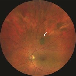
Image of a choroidal nevus.
A choroidal nevus is a pigment lesion. Nevi can be found anywhere in the body that has pigment, including skin, mucus membranes, internal organs, and of course, your eye. Nevi can be found in the front of the eye, in the conjunctiva, and in the iris, but are most commonly found under the retina in the choroid. Nevi vary in size and shape. They are not present at birth. Nevi remain flat and do not typically grow. They do not interfere with vision. Up to 5% of normal individuals can have choroidal nevi.

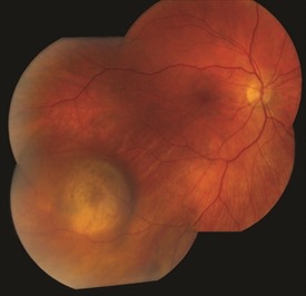
Image of a choroidal melanoma.
If a nevus starts to grow, it may develop into a melanoma. This process is rare, and occurs in about 1:10,000 cases. Just like skin melanoma, choroidal melanomas may spread to other part of the body and cause significant illness, and even death. Consequently, patients with choroidal nevi need to be monitored about once a year.
If a choroidal melanoma is small, it can be treated with radiation. This is done with a plaque filled with radioactive seeds. The plaque is surgically placed under the melanoma for a few days, and is then removed. Patients with ocular melanoma need to be monitored by both a retina specialist and an oncologist for evidence of recurrence, growth or spread elsewhere in the body.