Retinal Artery Occlusion

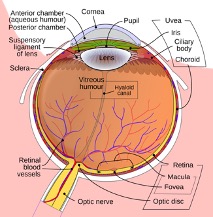
Illustration of ocular circulation.
Just like any other tissue in your body, the retina has a blood supply. The central retinal artery, travels down the optic nerve and supplies blood to the entire retina. At the point the central retinal artery enters the retina, it begins to progressively branch into smaller arteries and then microscopic capillaries, which supplies oxygen and nutrients to the retina. This arterial system connects to a parallel set of veins that drain into larger veins leading to the central retinal vein, which exits the retina also through the center of the optic nerve. Some of the most common problems a retina specialist sees are related to abnormalities in the blood vessel system of the retina.
A retinal artery occlusion is a blockage of either the central retinal artery of one of its branches. Since the retina develops along with the brain, an artery occlusion is like a stroke. Typically, there is sudden painless loss of vision in one eye with a defect in the field of vision. The retinal artery occlusion may be transient and last for only a few seconds or minutes if the blockage breaks up and restores blood flow to the retina, or it may be permanent. If you are lucky enough to be near a hospital or your doctor’s office, emergency treatment may help. In any event, you should seek medical attention as soon as possible.

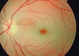
Image of a central artery occlusion.
There are two types of artery occlusions: arteritic and non-arteritic. Arteritic occlusions occur in individuals over the age of 65, and are associated with inflammation in arteries throughout the body. This is a dangerous situation and must be diagnosed and treated promptly. Laboratory tests and an artery biopsy confirm the diagnosis. The treatment is steroids. Non-arteritic occlusions occur due to narrowing of large arteries such as the carotid artery or valvular heart disease. In this case a small blood clot or embolus is released and blocks the retinal artery. Again, it is important to seek medical attention quickly to avoid complications such as a stroke or a heart attack.