Retinal Tears & Holes

The retina is the light sensitive tissue which lines the back of the eye and forms the optic nerve. Tears may occur in the retina with the risk of a retinal detachment and severe loss of vision. Retinal tears occur as a result of the process of posterior vitreous detachment. At birth, the vitreous has the consistency of Jello and is firmly adherent to the retina. Later in life, the vitreous begins to liquify and partially separate from the retina. At this point, patients may experience small floaters. As the vitreous shrinks and separates more completely from the retina, larger floaters may be seen. If the vitreous remains adherent to the retina, the patient may experience a light flash. As the vitreous contracts, it may pull on the retina with sufficient force to tear the retina. Since the retina contains many blood vessels, bleeding into the vitreous cavity may occur resulting in large floaters.

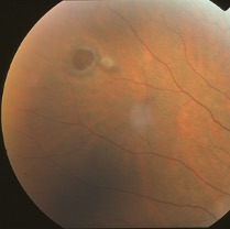
Image of a retinal hole.
Both retinal holes and retinal tears can lead to retinal detachment. This is typically an acute event associated with large floaters, light flashes and a progressive defect in vision, like a curtain. Should any of these symptoms occur, you should contact your optometrist or ophthalmologist who will determine whether you need to be seen by a retina specialist. Urgent care centers and emergency rooms are not equipped to determine whether a retinal detachment is present so you should be seen as soon as possible by your eye care provider.
The decision to treat a retina tear or hole depends on the clinical findings and risk factors for retinal detachment. Larger tears, associated with bleeding, are a high risk and should be treated. Retinal holes without symptoms can be safely followed. Patients with a high degree of myopia (nearsightedness), a family history of retinal detachment, previous cataract surgery or a tear or detachment in the fellow eye are at greater risk.
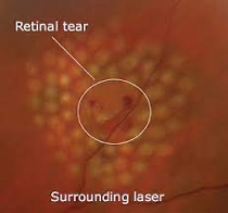
Retinal tears and holes are typically treated with laser. The procedure is done in the office with topical anesthesia using a contact lens. The laser spot-welds the retina around the retinal defect preventing the development of a detachment. Cryotherapy or freezing may also be used. There is a period of time after treatment, lasting about two months, where problems may develop. These would include new retinal tears or a retinal detachment. If you experience new large floaters, light flashes or a defect in your vision, you should be rechecked.
See Fact Sheet about Posterior Vitreous Detachment.*